
If you've never heard of your back teeth, you might be wondering what they are and how to brush them. This article will give you a general overview of your back teeth. Learn about the Central incisors, the Third molars, and the Premolars. These teeth are often difficult to brush, and they're more prone to decay and cavities. Fortunately, there are several ways to maintain a healthy set of back teeth.
Table of Contents
Canine teeth
The back teeth in dogs are called premolars. These teeth are located behind the canine teeth and are used to shear food. When your dog chews on the side of the mouth, he is using his premolars. The canines are the heavy-duty teeth in the back of your dog's mouth. They are the longest tooth roots and have a single, pointy cusp.
They are the longest and sharpest of the back teeth and are used to rip food apart. The two sets of canines are usually on opposite sides of the mouth. The canines come through first, and the lower one comes through before the upper. Premolars are the teeth that come before the molars. In addition to the two rows of premolars, dogs also have eight molars on opposite sides of their mouths.
A surgeon may use the periosteal elevator to lift a small portion of mucosa surrounding the tooth to expose the bone. If the bone remains attached to the tooth, normal healing should occur, but if the bone has broken away from the tooth, it will prolong the healing process. During extraction, the tooth should be stabilized with forceps before being extracted. The bone may be left in place, attached to the periosteum. When this is possible, the bone should be removed if it is nonvital.
A dentist can give you a report card and a detailed dental chart so that you can monitor the progress of your pet's dental health. If your dog has stopped eating, you should take your pet to the vet for an examination. Your veterinarian can help you prevent this from happening by discussing dental health during your pet's annual examination. You can also choose to have a dental cleaning if you suspect that your dog has periodontal disease.
The goal of dental treatment in children with CP is to preserve the deciduous teeth and avoid extraction. In cases where root resorption of the deciduous teeth occurs, the pediatric dentist will refer the child to a prosthodontist or orthodontist. CP is managed with a multidisciplinary team, including a pediatric dentist who manages the child's behavior, performs intermediate restorations, and treats malocclusion.
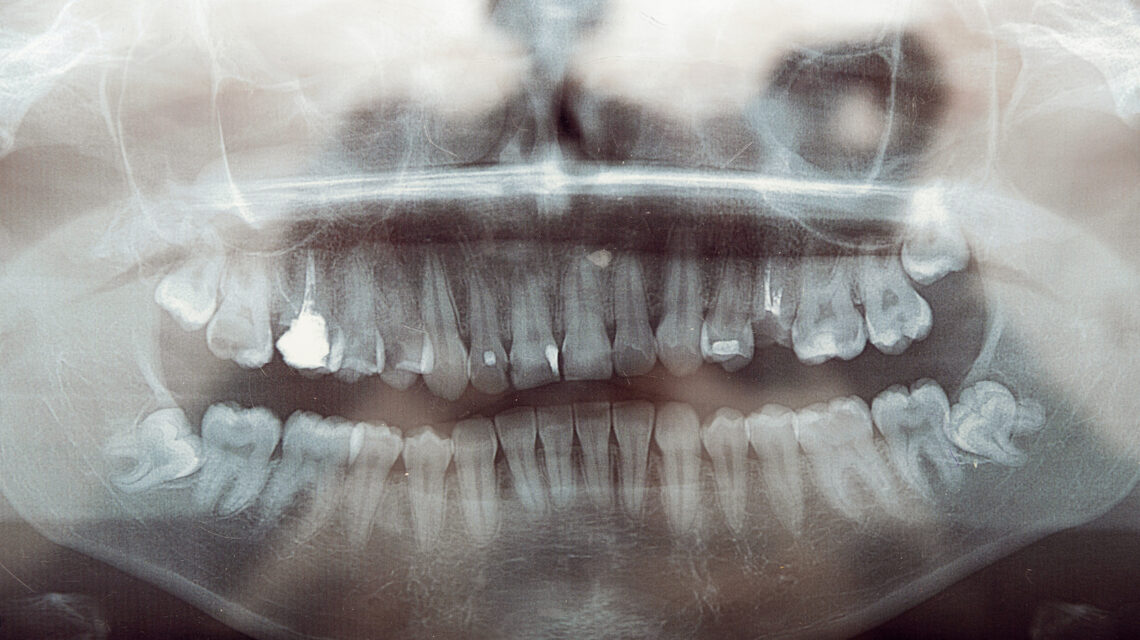
Third molars
The third molars are the oldest teeth in the human mouth and have many important functions. However, their eruption into the mouth can be painful and can even cause cavities. Additionally, the teeth in front of these molars are at risk of shifting and developing crookedness, which can lead to further problems. If you suspect that you are experiencing this, it's best to see your dentist for a consultation. You can use the Healthline FindCare tool to find a dentist in your area.
In Scotland, a recent survey of dental services shows a 7% increase in activity for third molars. The trend is similar to the upward trend of activity in general dental care in Scotland and England. In Scotland, the number of day-case/in-patient third molar procedures increased by 67%, and the overall number of third molars removed grew by 130%. The number of patients undergoing these procedures also increased.
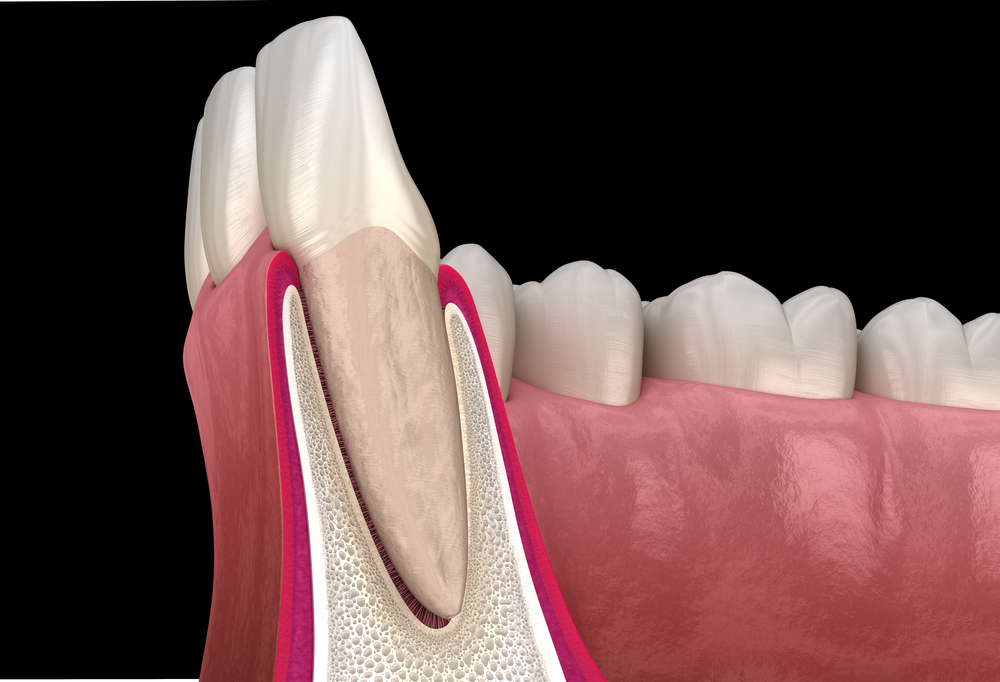
The removal of third molars is one of the most common procedures in oral surgery, with around 10 million teeth extracted every year. This is often required for various reasons, including the risk of impaction associated with caries, periodontal defects on the distal surface of the second molars, and odontogenic cysts. Furthermore, while a majority of general dentists recommend third molar extraction, the research does not show a clear pattern of third molar eruption. This is because third molars are rare and are not completely erupted.
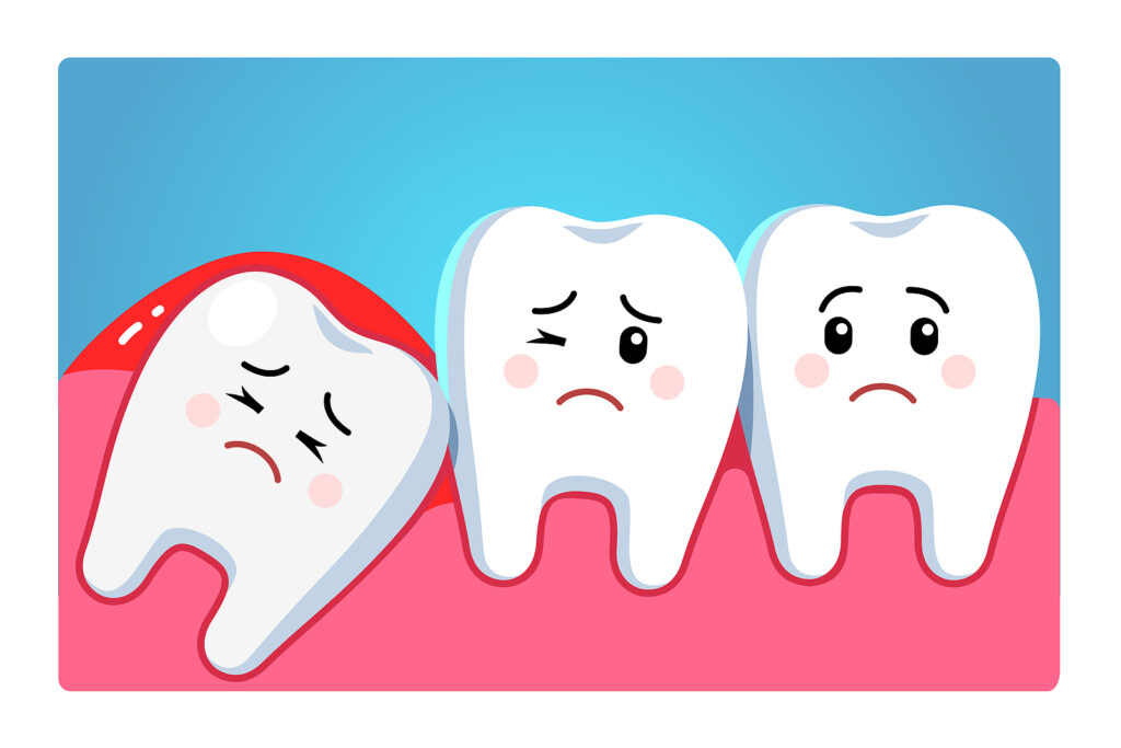
When the third molars become impacted in the mouth, it can cause considerable pain and misalignment of the teeth. An impacted third molar is likely to irritate the surrounding teeth and may be the cause of an oral infection. If the third molars are causing significant discomfort, it is best to have them extracted to maintain optimal oral health. If not, it can lead to more complications.
Among the teeth, the third molars are also known as wisdom teeth. Though third molars are beneficial for chewing, the wrong eruption can cause damage to the other teeth. If your wisdom teeth are impacted, visit your dentist for a professional evaluation. These teeth are typically the last to appear and can pose a serious risk to your oral health. Therefore, it's crucial to seek professional advice if you're experiencing any pain.
Central incisors
The central incisors are the first teeth to erupt in the mouth. They serve essential functions in everyday life, including chewing and maintaining the position of the jaw. As such, they need to be strong and clean. Proper cleaning and regular oral care will help you keep these teeth in top shape and function efficiently. Whether you have one or two, remember to talk to your dentist about any urgent dental issues.
The distal surface of the central incisor has a cervical line and a deeply marked central groove. Its mesiodistal and lingual surfaces are similar, but the incisal edge is broader. This tooth has a cingulum (the area behind the incisal edge), and a straight outline. The distal edge of the central incisor is similar to that of the lateral incisors.
The incisal ridge of a newly erupted incisor is formed by three small mamelons that wear down to a flat edge. The mandibular central incisor is almost symmetrical in anatomy. Its proximal and labial surfaces are triangular, and the incisal ridge is triangular. Some incisors also have vertical developmental depressions that provide anchorage for the tooth.
The pulp chamber of a central incisor resembles the pulp chamber of the lower central incisor. The mesiodistal portion of the root resembles a cone with a rounded apex. The lateral incisors have a longer, more conical root than the central incisors. The crown is wider than the incisal portion of the central incisor.
While the first two molars appear early in life, the central incisors will be the last to erupt. The first four permanent teeth will come through the gums behind the last baby tooth. The second set of permanent teeth, called wisdom teeth, will erupt in the late teens and early twenties. This will bring the total number of permanent teeth to 32. This is when you will experience the most noticeable changes in your smile and teeth.
The mesiodistal and lingual surfaces of central incisors are similar, but the distal surface is wider and more convex. The cervical line is curved and slightly offset distally. The incisal ridge has an incisal ridge centered lingually. In addition, the distal surface is slightly longer than the mesial surface, which gives the impression of a bigger tooth.
Premolars
The premolars are the back teeth, which serve a dual function in chewing and esthetics. They function much like the canines in tearing and grinding food, but have a slightly different anatomic form. Premolars have angular facial cusps, while the lingual cusps of the maxillary premolars are round. They have a complex series of curves on the occlusal surface, which must be kept healthy for life.
The purpose of premolars is to break up food and blend it with saliva for easier digestion. The eight premolars make up the complete dentition. The first and second premolars are paired and are located between the canines and the molars. The first one is the most prominent, with two cusps at its crown. The second premolars are located behind the first one. The secondary premolars have two or three cusps and are often found in groups of two teeth.
In addition to their position on the front teeth, the premolars are important for chewing food. In addition to the canines, these teeth serve as anchors for the other teeth. Children's permanent lower canines appear between age nine and twelve, with the upper canines emerging a little before the lower ones. Premolars are much larger than incisors, and have multiple ridges. They also help chew food and are responsible for the emergence of the molars, the last set of teeth.
The premolars are transitional teeth. They appear between the canines and the molars, and have at least two cusps. During chewing, the premolars grind up food and prepare it for digestion by the molars. The second set of premolars appears between ten and twelve years old. In general, the molars come in after the premolars, but are not present in infants.
The third set of teeth, the molars, are located in the back of the mouth. These teeth are also known as wisdom teeth. The third set of teeth, the third molar, erupts after the second set of premolars. These teeth are usually fused. A common way to remove wisdom teeth is surgical removal. In many cases, it is the best option. Despite their appearance, they can cause problems if left untreated.